Biology SKELETAL MUSCLE IN DETAIL

KEY TOPICS

`star` Components of Skeletal Mucles
`star` Sarcomere
`star` Structure of Contractile Proteins

COMPONENTS OF SKELETAL MUSCLES

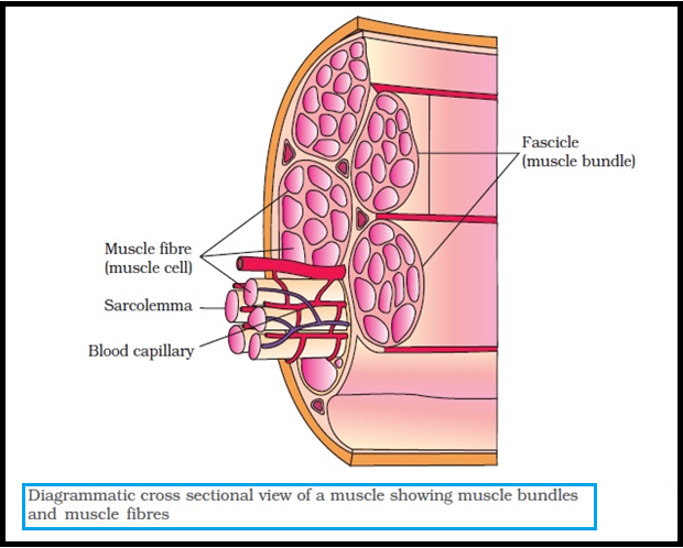
● Each `color{violet}("organised skeletal muscle")` in our body is made of a number of `color{brown}("muscle bundles")` or `color{brown}("fascicles")` held together by a `color{violet}("common collagenous")` connective tissue layer called `color{brown}("fascia.")`

● Each `color{violet}("muscle bundle")` contains a number of `color{brown}("muscle fibres.")`

● Each `color{violet}("muscle fibre")` is lined by the `color{violet}("plasma membrane")` called `color{brown}("sarcolemma")` enclosing the `color{violet}("sarcoplasm.")`

● `color{violet}("Muscle fibre")` is a `color{brown}("syncitium")` as the `color{violet}("sarcoplasm")` contains many `color{violet}("nuclei.")`

● The endoplasmic reticulum, i.e., `color{brown}("sarcoplasmic reticulum")` of the `color{violet}("muscle fibres")` is the store house of `color{violet}("calcium ions.")`

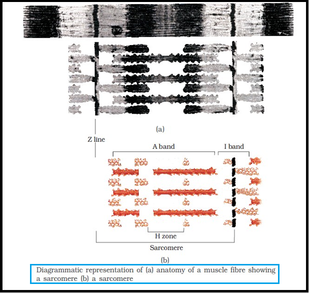
● A characteristic feature of the `color{violet}("muscle fibre")` is the presence of a large number of parallelly arranged filaments in the `color{violet}("sarcoplasm")` called myofilaments or `color{brown}("myofibrils.")`

● Each `color{violet}("myofibril")` has alternate `color{brown}("dark and light bands")` on it.

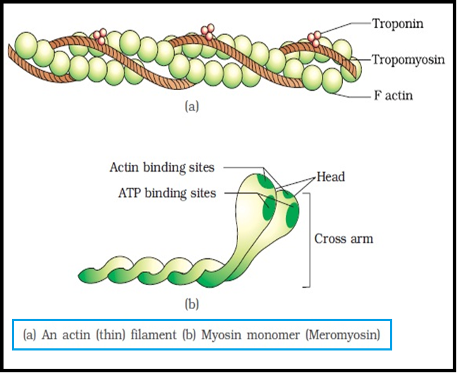
● A detailed study of the `color{violet}("myofibril")` has established that the striated appearance is due to the distribution pattern of two important proteins – `color{brown}("Actin and Myosin.")`

SARCOMERE

● The `color{violet}("light bands")` contain actin and is called `color{brown}("I-band or Isotropic band")`, whereas the `color{violet}("dark band")` called `color{brown}("‘A’ or Anisotropic")` band contains `color{violet}("myosin.")`

● Both the proteins are arranged as `color{violet}("rod-like structures, parallel")` to each other and also to the `color{violet}("longitudinal axis of the myofibrils.")`

● Actin filaments are thinner as compared to the `color{violet}("myosin filaments,")` hence are commonly called `color{brown}("thin and thick filaments")` respectively.

● In the centre of each `color{violet}("‘I’")` band is an elastic fibre called `color{brown}("‘Z’")` line which bisects it.

● The `color{violet}("thin filaments")` are firmly attached to the `color{violet}("‘Z’ line.")`

● The `color{violet}("thick filaments")` in the `color{violet}("‘A’ band")` are also held together in the middle of this band by a thin fibrous membrane called `color{brown}("‘M’ line.")`

● The `color{violet}("‘A’")` and `color{violet}("‘I’")` bands are arranged alternately throughout the length of the `color{violet}("myofibrils.")`

● The portion of the `color{violet}("myofibril")` between `color{brown}("two successive ‘Z’ lines")` is considered as the functional unit of contraction and is called a `color{brown}("sarcomere.")`

● In a `color{brown}("resting state,")` the edges of `color{violet}("thin filaments")` on either side of the `color{violet}("thick filaments")` partially overlap the free ends of the `color{violet}("thick filaments")` leaving the central part of the thick filaments.

● This central part of `color{violet}("thick filament,")` not overlapped by `color{violet}("thin filaments")` is called the `color{brown}("‘H’ zone")`.

STRUCTURE OF CONTRACTILE PROTEINS

`star` `color{green}("ACTIN")`

● Each actin `color{violet}("(thin) filament")` is made of two `color{brown}("‘F’ (filamentous) actins")` helically wound to each other.

● Each `color{violet}("‘F’ actin")` is a polymer of monomeric `color{brown}("‘G’ (Globular) actins.")`

● `color{violet}("Two filaments")` of another protein, `color{brown}("tropomyosin")` also run close to the `color{violet}("‘F’ actins")` throughout its length.

● A complex protein `color{brown}("Troponin")` is distributed at regular intervals on the `color{violet}("tropomyosin. ")`

● In the resting state a `color{violet}("subunit of troponin")` masks the active `color{violet}("binding sites")` for `color{violet}("myosin")` on the actin filaments.

`star` `color{green}("MYOSIN")`

● Each `color{violet}("myosin (thick) filament")` is also a `color{violet}("polymerised protein.")`

● Many `color{violet}("monomeric proteins")` called `color{brown}("Meromyosins")` constitute one `color{violet}("thick filament.")`

● Each `color{violet}("meromyosin")` has two important parts, a `color{violet}("globular head ")` with a short arm and a tail, the former being called the `color{brown}("heavy meromyosin (HMM)")` and the latter, the `color{brown}("light meromyosin (LMM).")`

● The `color{violet}("HMM component")`, i.e.; the head and short arm projects outwards at `color{violet}("regular distance")` and angle from each other from the surface of a `color{violet}("polymerised myosin filament")` and is known as `color{brown}("cross arm.")`

● The `color{violet}("globular head")` is an active `color{brown}("ATPase enzyme")` and has binding sites for `color{violet}("ATP")` and active sites for `color{violet}("actin.")`


SiteLock